


The Art in Science competition invites artists, students and anyone interested in visual creativity to reinterpret images created during research at the ELI facilities. By adding colour, texture and graphic elements, participants can transform scientific images into original works of art.
The challenge is simple: Choose an image, keep its original lines and shapes, and make it your own through colour, graphic enhancement and any technique you like: paint, pencil, collage, textile, digital art or something we haven't imagined yet. The 12 most popular works will feature in the ELI 2027 wall calendar, shared across both facilities.
The images to be reinterpreted:
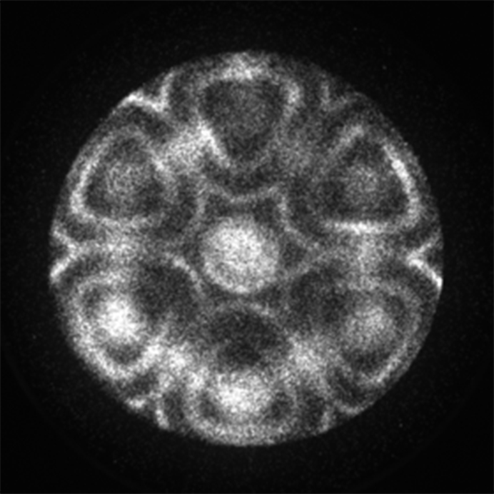
Image captured by the NanoESCA Group (ELI ALPS). Electron flower. The image shows a momentum-space map of the electronic structure of a WSe₂ sample covered with a molecular FePc layer, acquired using a photoemission electron microscope (NanoESCA). During the measurement, the emitted electrons were imaged according to their momentum rather than their spatial position on the sample.
The intensity and momentum distribution of the electronic states evolve continuously in the energy-resolved slices recorded at different kinetic energies. At certain energies, the distribution forms a striking pattern resembling the petals of a flower. The image therefore does not represent the real-space surface morphology of the sample, but rather the intensity distribution formed in the reciprocal, or momentum, space of the emitted electrons. The regular patterns are likely determined by the electronic structure and crystal symmetry of the material, together with contributions from the electronic states of WSe₂ and the evaporated FePc molecular layer.
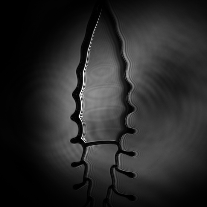
Image credit: Indranuj Dey (ELI ALPS). The image captures a particular instant during the disconnection of a liquid leaf target exhibiting complex fluid dynamics before detachment of multiple droplets. Captured using a scientific CMOS camera in the SYLOS SHHG beamline.
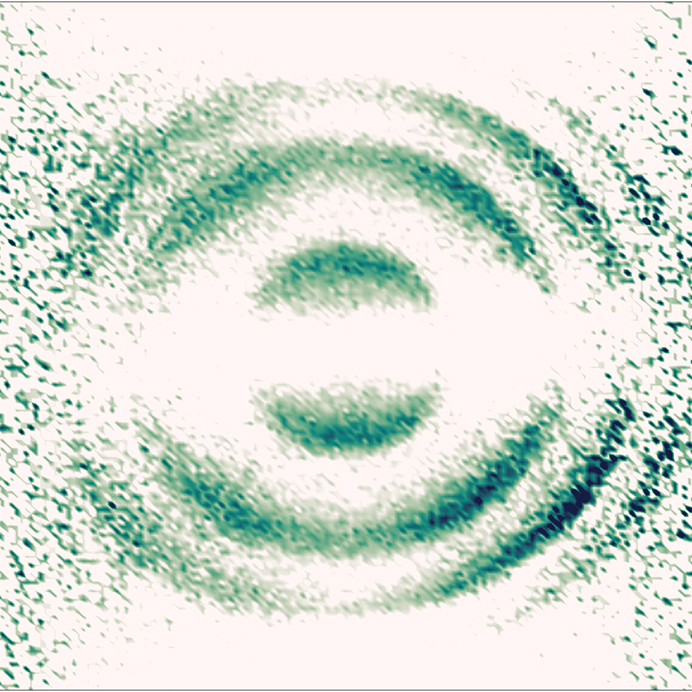
Image credit: Tamás Csizmadia (ELI ALPS). Angular distribution of photoelectrons from the ionization of helium. This image was recorded in the LTA4 laboratory using a reaction microscope end-station. It depicts the angle-resolved momentum distribution of photoelectrons from the photoionization of helium by extreme ultraviolet harmonics. It was captured in the polarization plane of the ionizing radiation.
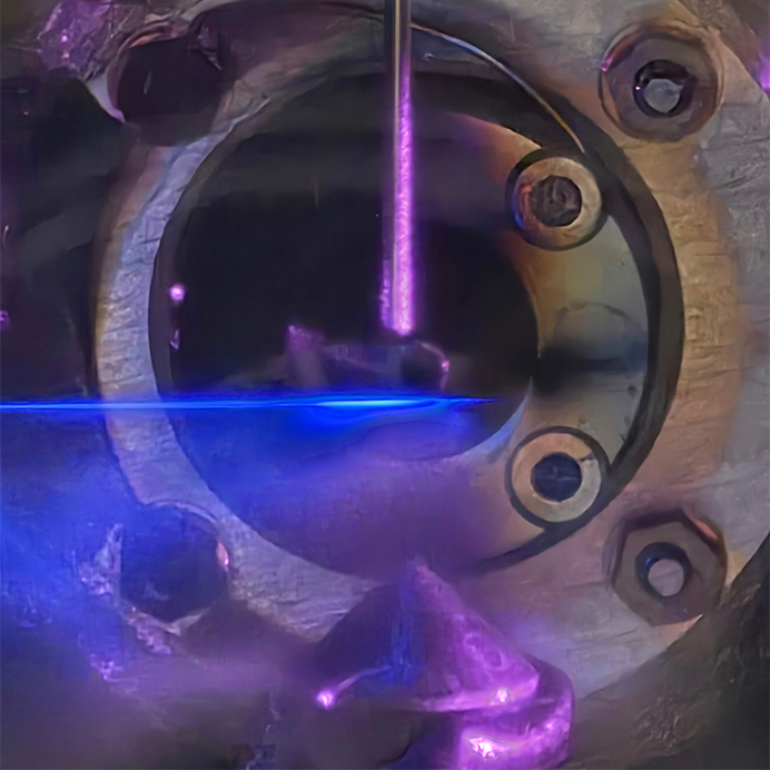
Image credit: Rohit Kumar (ELI ALPS). Plasma beam created by the laser in air in front of the time-of-flight spectrometer.
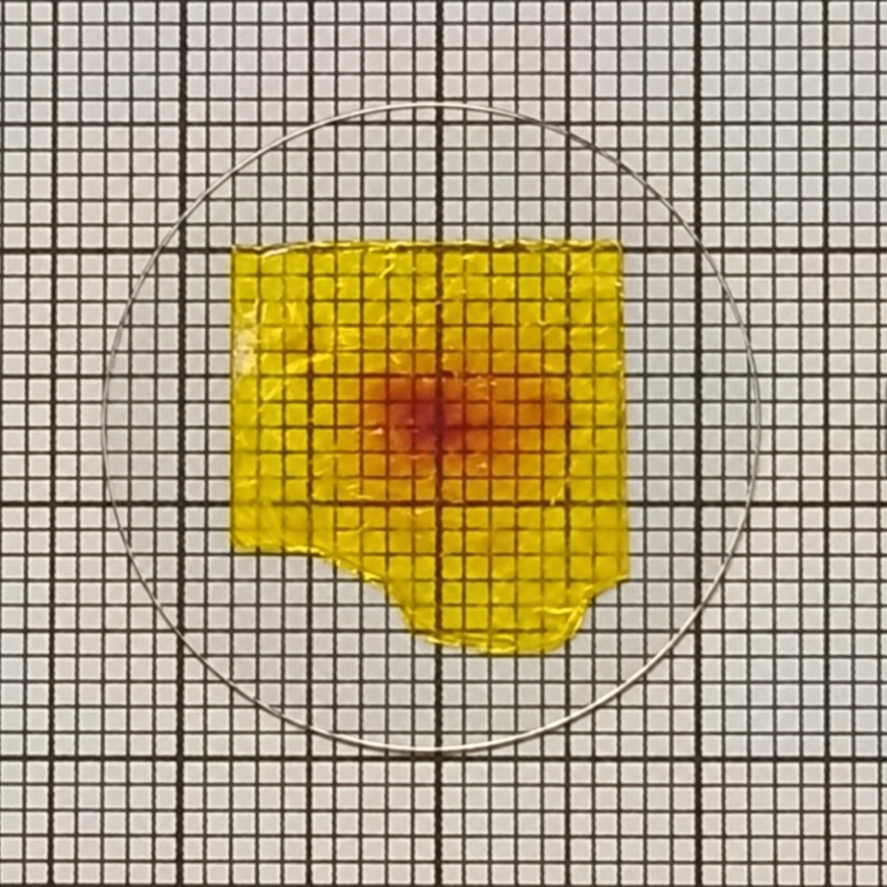
Image credit: Abhishek Gupta, Tianmiao Zhang (ELI ALPS). Photograph of large organic molecular crystals of different compositions, fused onto round sapphire plates for improved heat conductivity. The crystals are used for producing terahertz-frequency radiation from a laser beam.
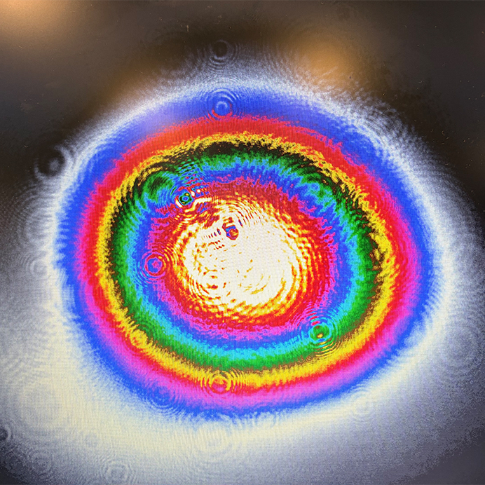
Image credit: Krisztina Trényi-Sárosi & Imre Seres (ELI ALPS). The image was taken in the HR-1 laboratory at ELI ALPS and shows the beam profile of an ultrashort laser pulse before it enters the transient absorption spectroscopy endstation. The colours represent different intensity levels. The symmetric, centrally concentrated profile indicates good (Gaussian) beam quality and proper optical alignment. The aligned pulses are then used to follow ultrafast processes in materials on timescales ranging from femtoseconds to nanoseconds.
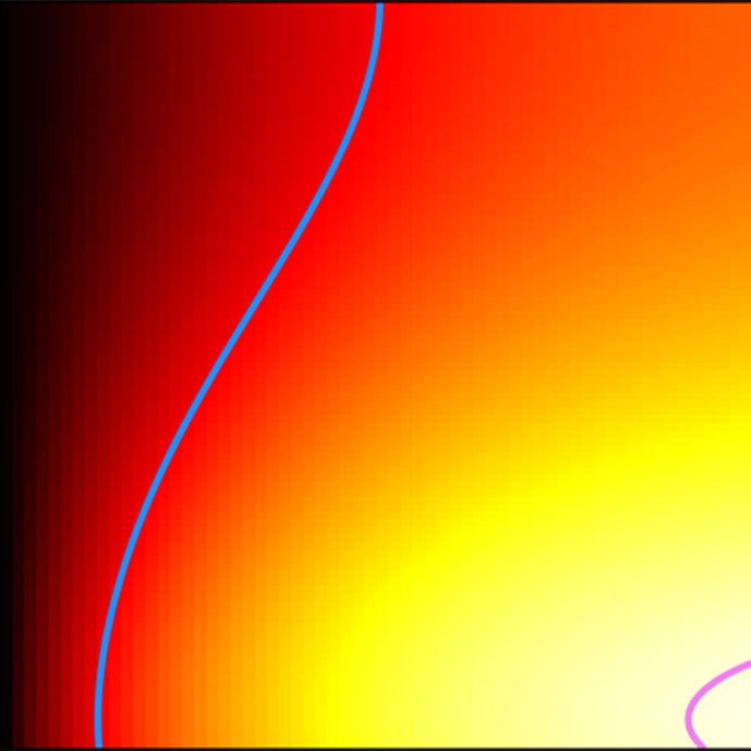
Image credit: Mousumi Upadhyay Kahaly (ELI ALPS). This is from the simulation of laser induced heating in gold samples produced by a short 12.5 fs pulse.
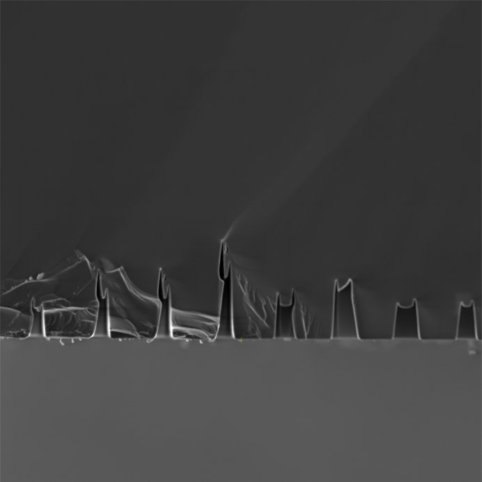
Image credit: Judit Budai and Zsuzsanna Márton (ELI ALPS). Scanning electronmicroscope image of the cross section of focussed ion beam drilled holes in crystalline silicon.
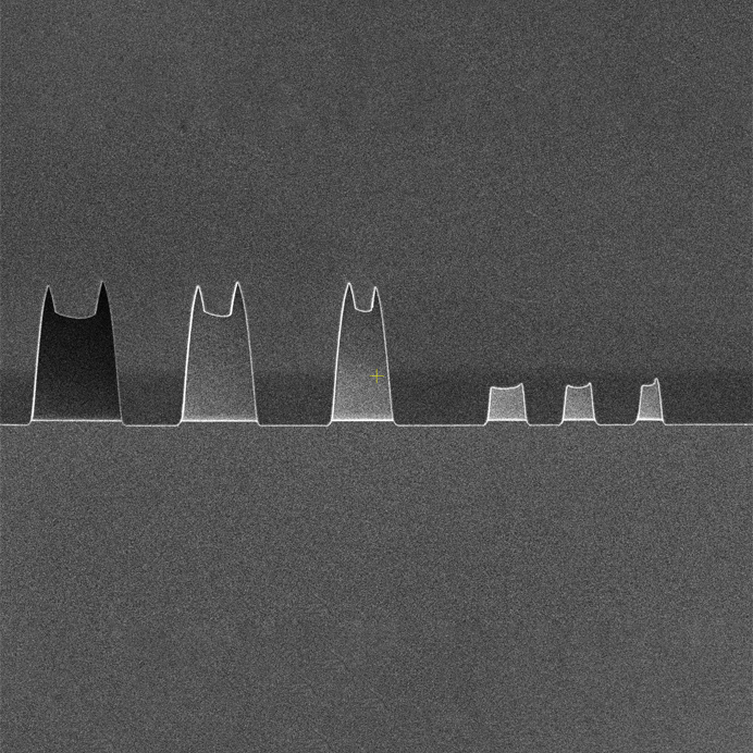
Image credit: Judit Budai and Zsuzsanna Márton (ELI ALPS). Scanning electronmicroscope image of the cross section of focussed ion beam drilled holes in crystalline silicon.
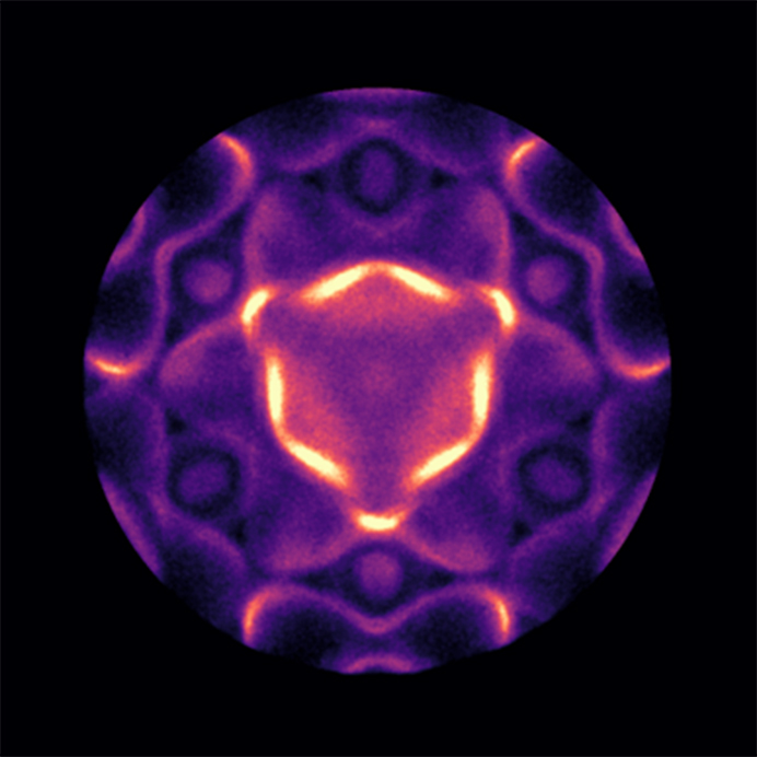
Image captured by the NanoESCA Group (ELI ALPS). Determination of the electronic structure near the Fermi level of a gold single crystal (Ir111), i.e., an inverse field mapping of a constant-energy slice at a kinetic energy of 21.1 eV, the highest charged energy level. A so-called electron band structure recording made in impulse, also known as momentum space mode, with a photoemission electron microscope. Only the valence electrons with low binding energy are ejected from the sample as a result of the low excitation photon energy, and we owe this momentum-space structure itself to this ordered 111 crystalline structure.
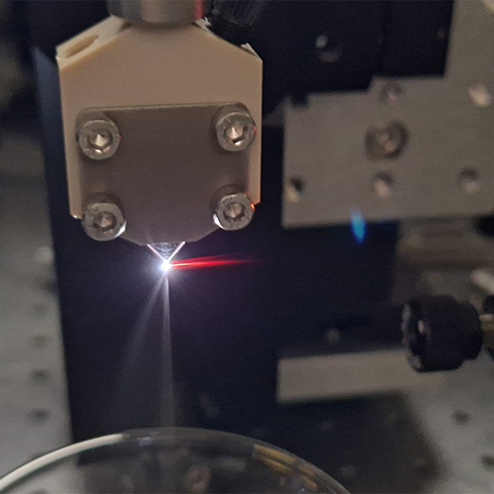
Image credit: Balázs Farkas (ELI ALPS). The birth of light. Plasma and high harmonic generation in liquid water using high intensity mid-infrared laser light. The goal is to study the structure of liquid water with the high harmonic generation process.
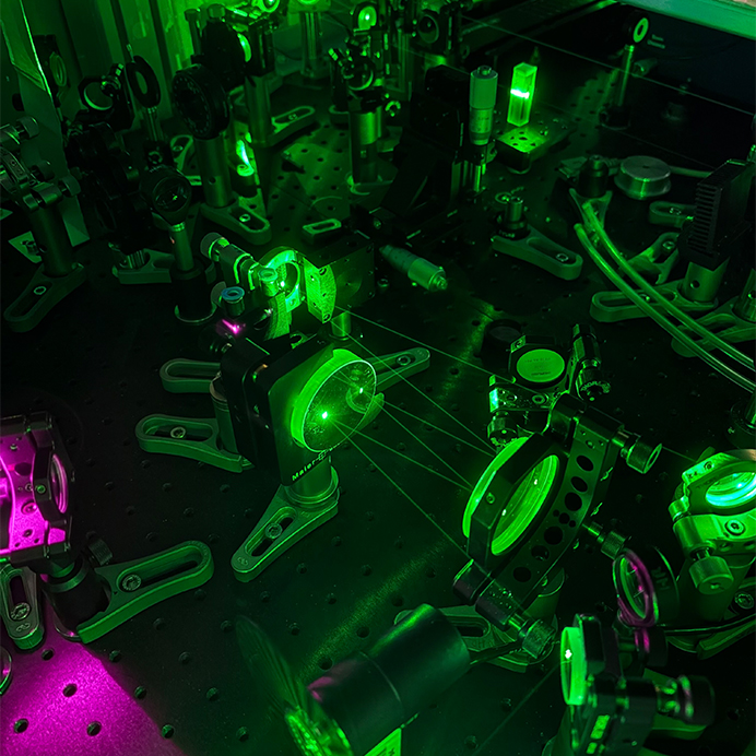
Image credit: Krisztina Trényi-Sárosi (ELI ALPS). The image was taken in the HR-1 laboratory at ELI ALPS and shows the chirped mirrors of the transient absorption spectroscopy setup. The laser beam path is also clearly visible. Different wavelengths are reflected from different depths inside the mirrors, giving them different delays. This helps compensate for dispersion and keeps the laser pulses short, which is important for femtosecond measurements.
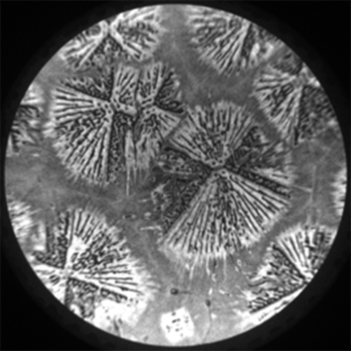
Image captured by the NanoESCA Group (ELI ALPS). The image shows BAPI perovskite single crystals grown on an ITO substrate and recorded using photoemission electron microscopy (PEEM). The image was acquired in real-space mode with a 300 µm field of view, revealing the characteristic radial morphology of the single crystals. The image contrast is based on the local work function, making the selected area suitable for mapping the electronic band structure of the investigated surface.
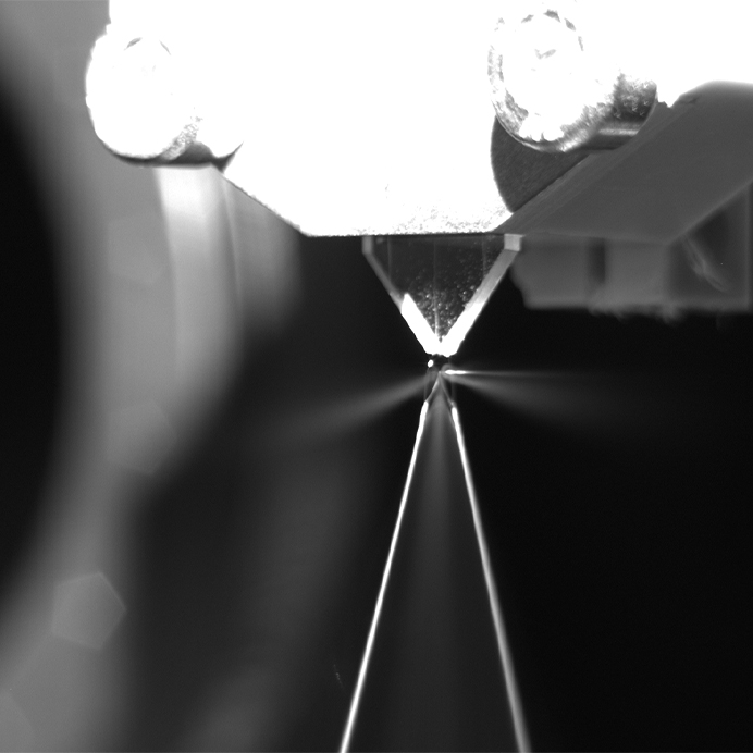
Image credit: Levente Ábrók (ELI ALPS). High harmonic generation in liquid jet
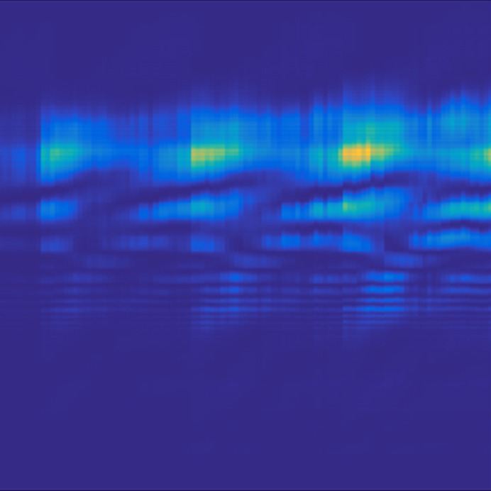
Image credit: Levente Ábrók (ELI ALPS). Carrier-envelope phase dependence of high harmonics generated in ZnO crystals.
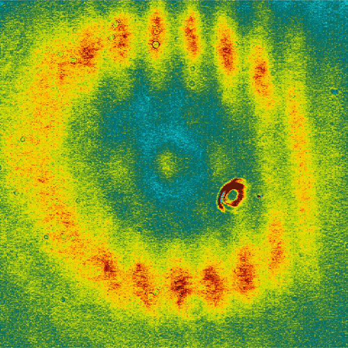
Image credit: Levente Ábrók (ELI ALPS). Time zero setting of a pump-probe setup using the MIR beam.
The images captured at the ELI facilities can be downloaded in high resolution from here.
Application requirements:
The completed works can be submitted either on paper or in digital format. An applicant may submit a maximum of 2 works.
Technical requirements:
• square-shaped artwork
• minimum size: 20 × 20 cm
Additional technical requirements for works created with digital techniques:
• CMYK settings
• 300 dpi resolution
• maximum file size of 15 MB
The following must be enclosed with the application:
• title of the artwork
• applicant's name
• applicant's age
• applicant's place of residence (city, country)
• if relevant: name of school, city and country, grade, name of the preparing teacher
• applicant's email address (in the absence of an email address, we will not be able to notify applicants of the results)
The deadline for submitting entries is November 8, 2026.
Digital works can be sent to artinscience@eli-alps.hu.
Paper-based artworks are welcome in person or by mail at either of the two institutions; please write on the envelope: "Art in Science competition".
ELI ALPS Facility
Wolfgang Sandner utca 3.
6728 Szeged
Hungary
ELI Beamlines Facility
Za Radnicí 835
252 41 Dolní Břežany
Czech Republic
The entries will be evaluated jointly by the staff of ELI ALPS and ELI Beamlines. The 12 entries receiving the most votes will be included in the ELI 2027 wall calendar, and the winners will receive gifts. Applicants will be notified of the results and the details of the award ceremony by email.