


Similar to earlier competitions, entries must be the reinterpretation of images taken at our research institute. Contestants must retain the lines and forms of the original images but must recreate them by adding colour and using a technique of their choice. The finished works may be submitted on paper or in digital format. Each artist may submit up to two entries.
The images to be reinterpreted:
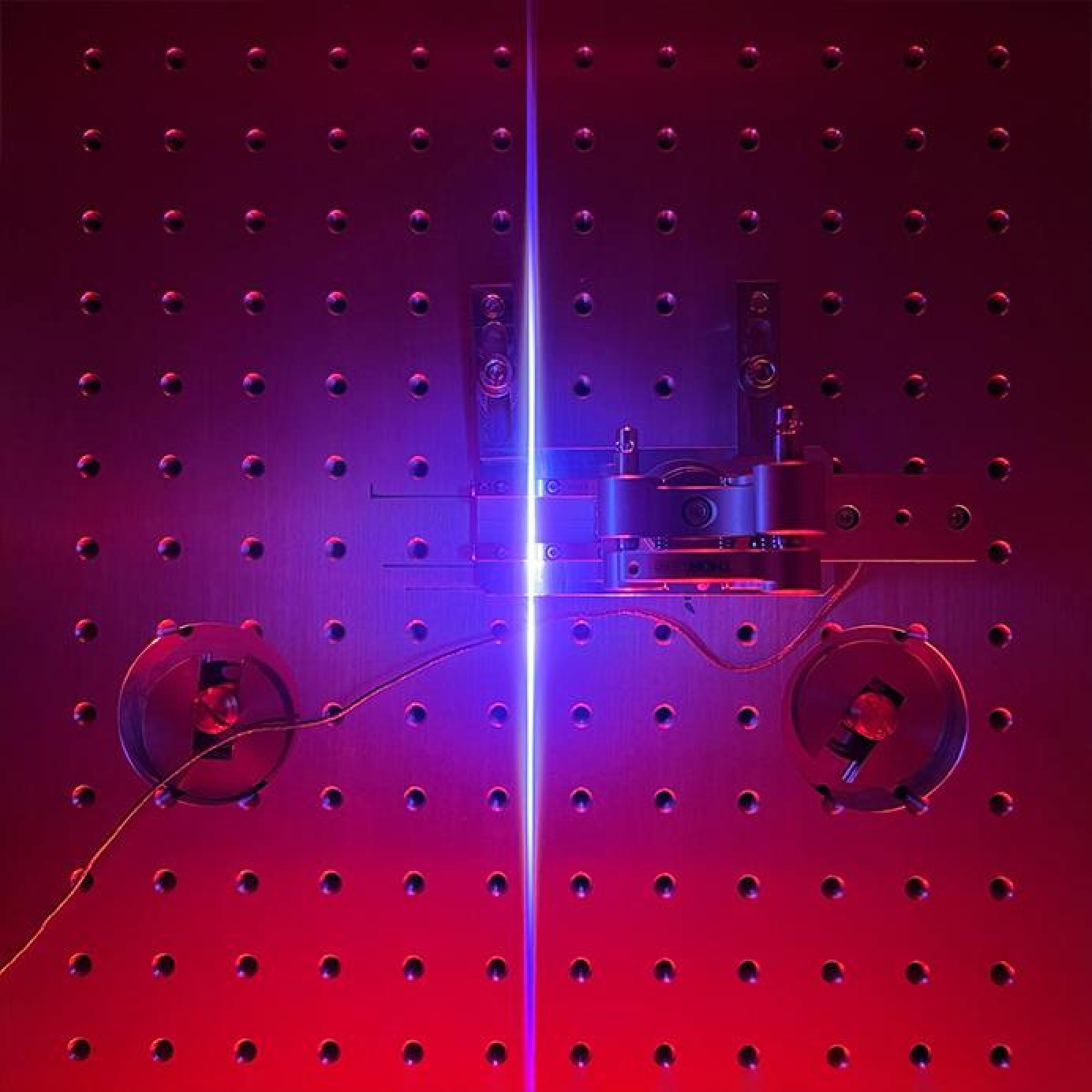
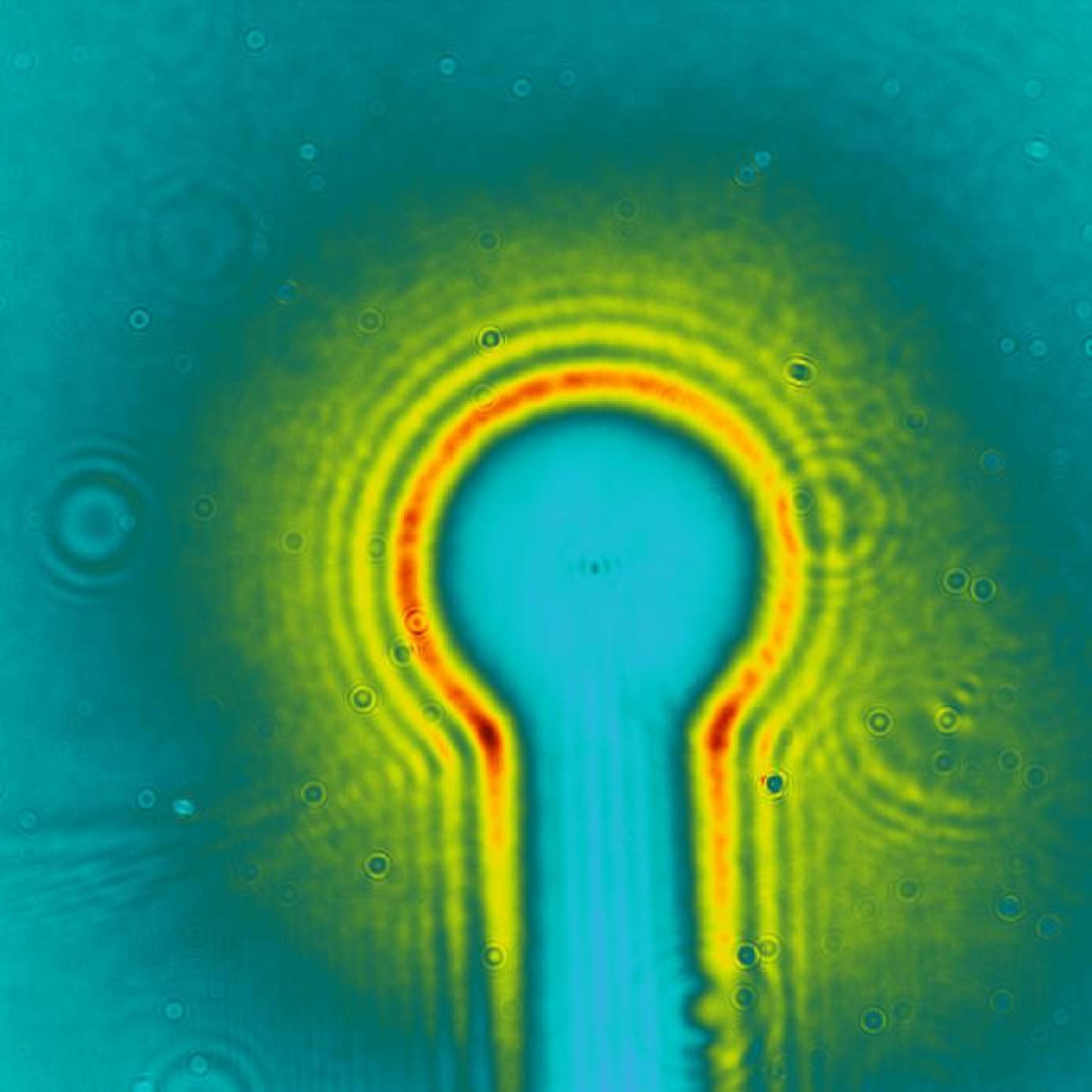
A significant milestone was recently achieved in the MTA laboratory with the SYLOS 3 laser system. The aim of the experiment was to further shorten the already ultrashort pulses of SYLOS 3 using the so-called post-compression technique. Several test runs were conducted, during which not only quantitative measurements were taken, but also vivid photographs were captured—highlighting the visual beauty of this colorful experiment. Inside the experimental chamber, when the focused, full-power SYLOS 3 pulses entered a low-pressure air environment, a bright, thin blue line appeared due to the scattering of ionization light—an impressive visual manifestation of the process.
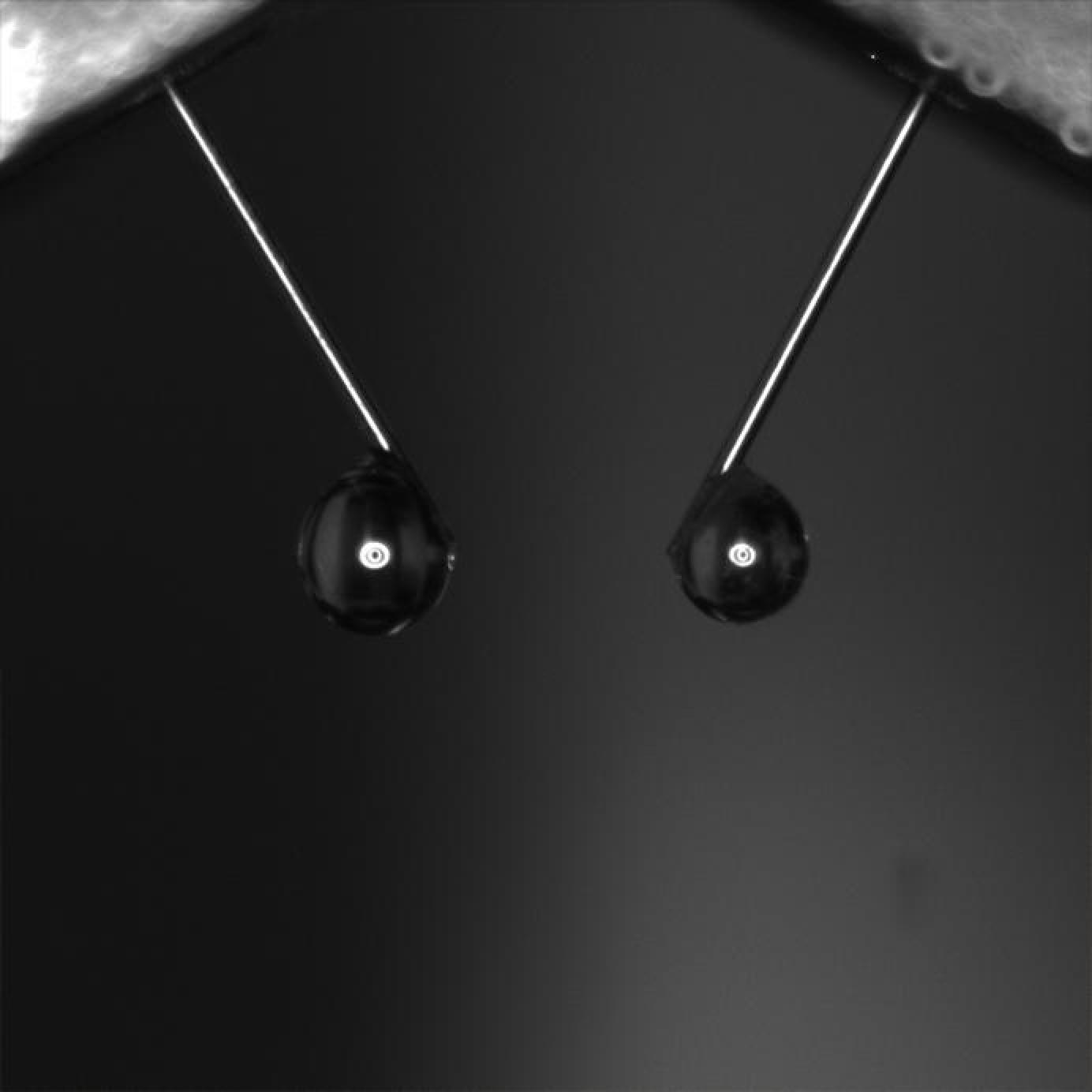
The image was captured by a CMOS camera with an imaging lens attached. The image depicts how ethylene glycol droplets roll up the polyamide cladding of the fused silica capillaries due to surface tension and form millimetre sized droplets, before dropping off.
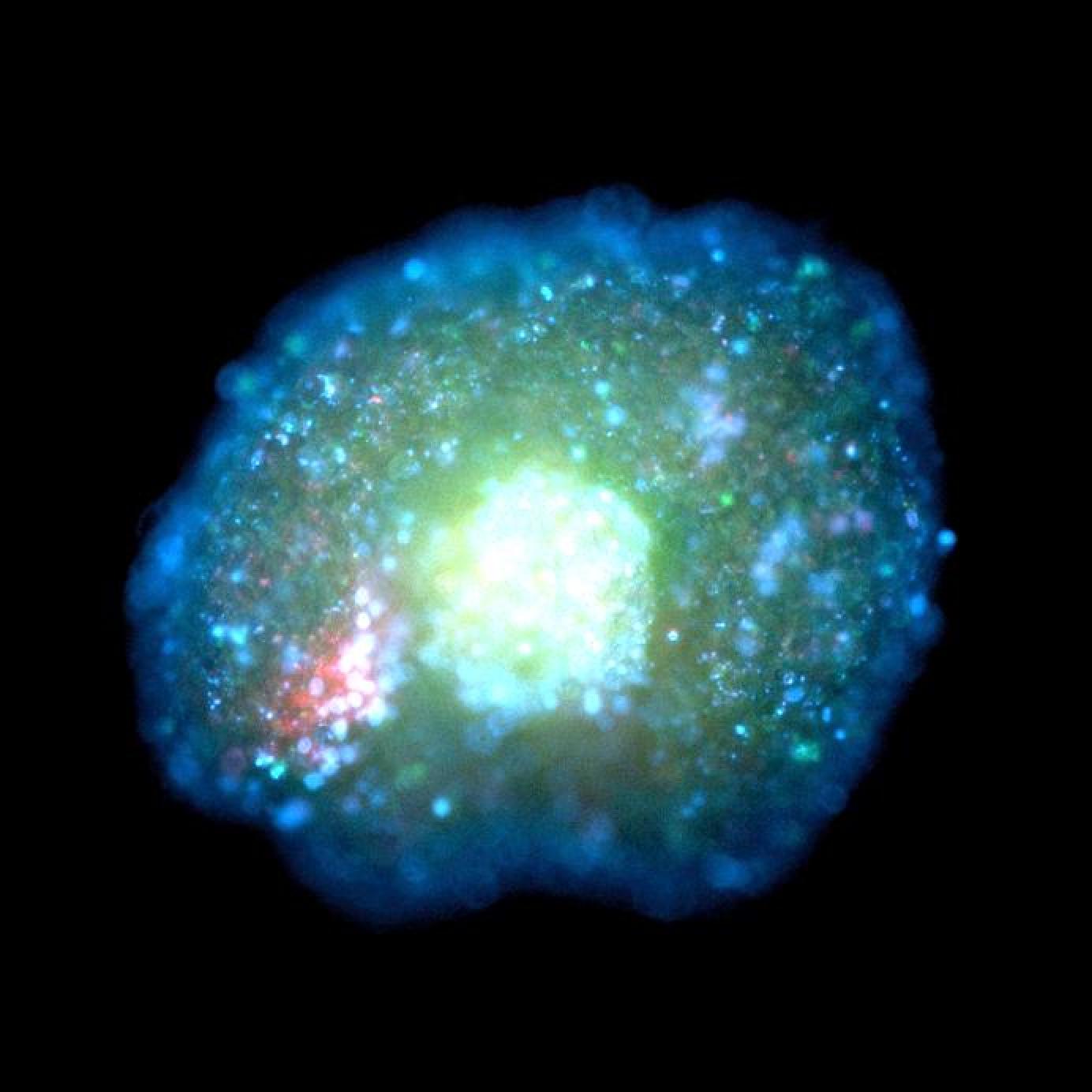
Image captured by the Biomedical Applications Research Group. The image shows a three-dimensional co-culture of normal human keratinocytes and fibroblasts. The cell nuclei were stained with blue Hoechst 33342 fluorescent nuclear stain; the green colour indicates programmed cell death (apoptosis), while red indicates necrotic cell death. The image was taken with a Zeiss Axio Observer Z1 fluorescence microscope at 10x magnification.
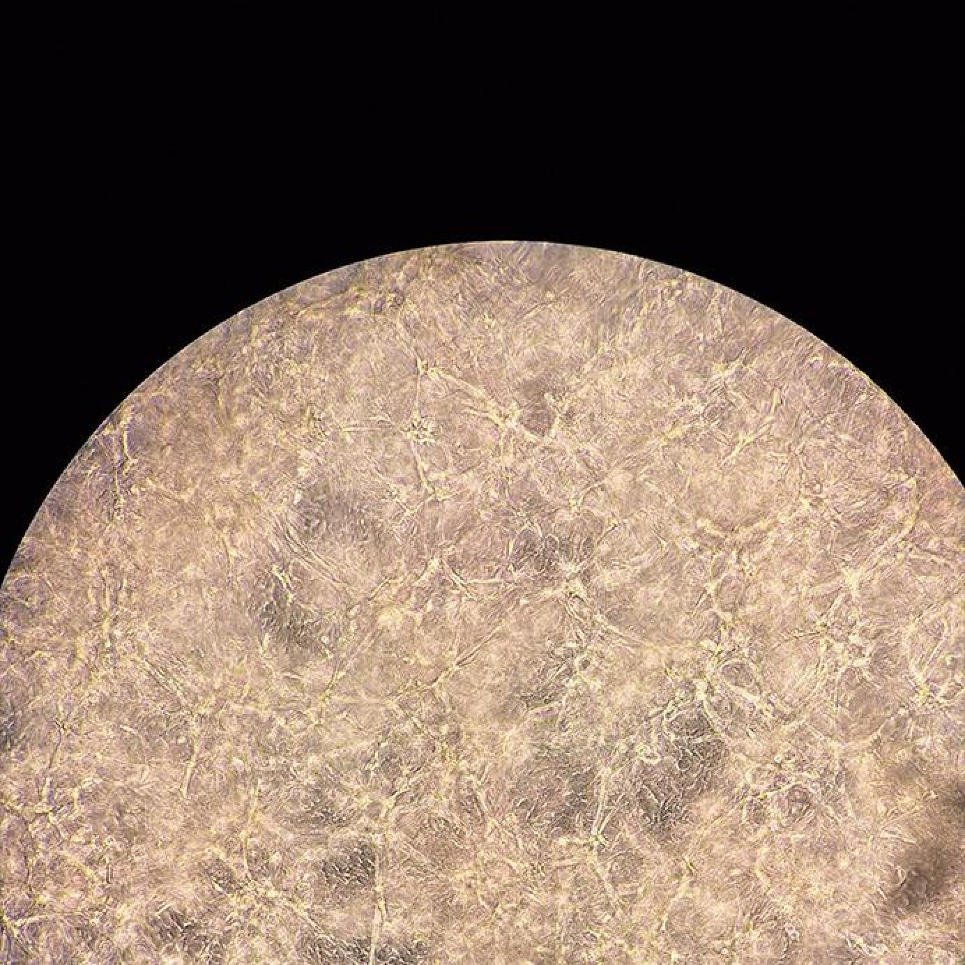
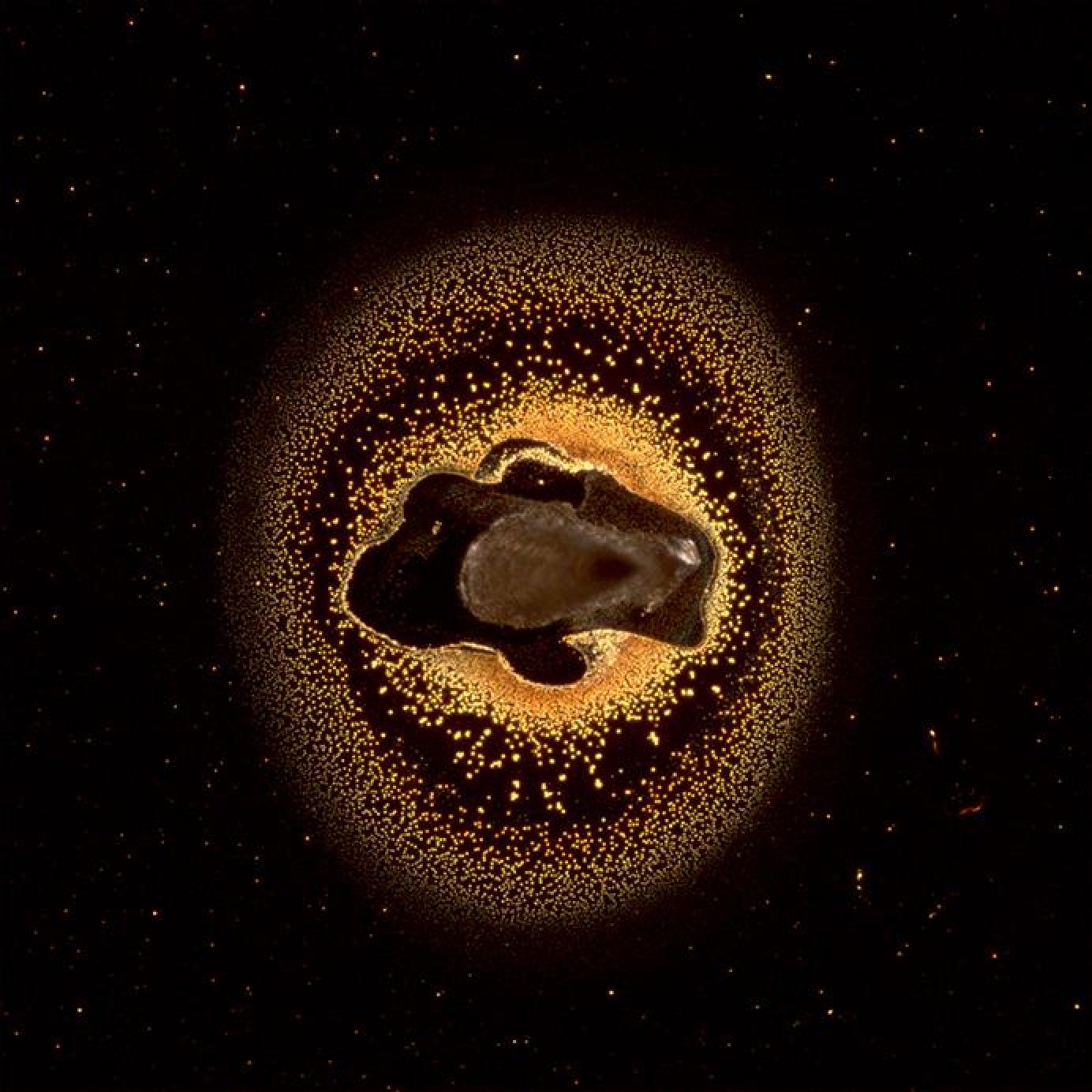
Image captured by the Biomedical Applications Research Group. The image shows three-dimensional cell connections created in Matrigel from human glioblastoma multiforme cells. The image was captured using a Euromex Oxion Inverso phase contrast microscope at 20x magnification.
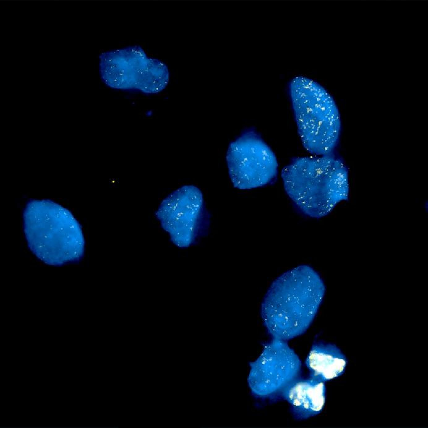
Image produced by the Biomedical Applications Research Group. The image shows the blue-stained nuclei of human glioblastoma multiforme cells, with the yellow dots indicating DNA double-strand breaks caused by 2 Gy irradiation. The almost completely yellow cells in the lower right corner indicate dividing cells. The cell nuclei were stained with blue Hoechst 33342 fluorescent nuclear stain, and the DNA double-strand breaks were stained with Alexa Fluor 488 fluorescently labelled gH2AX antibody. The image was taken with a Zeiss Axio Observer Z1 fluorescence microscope at 40x magnification.
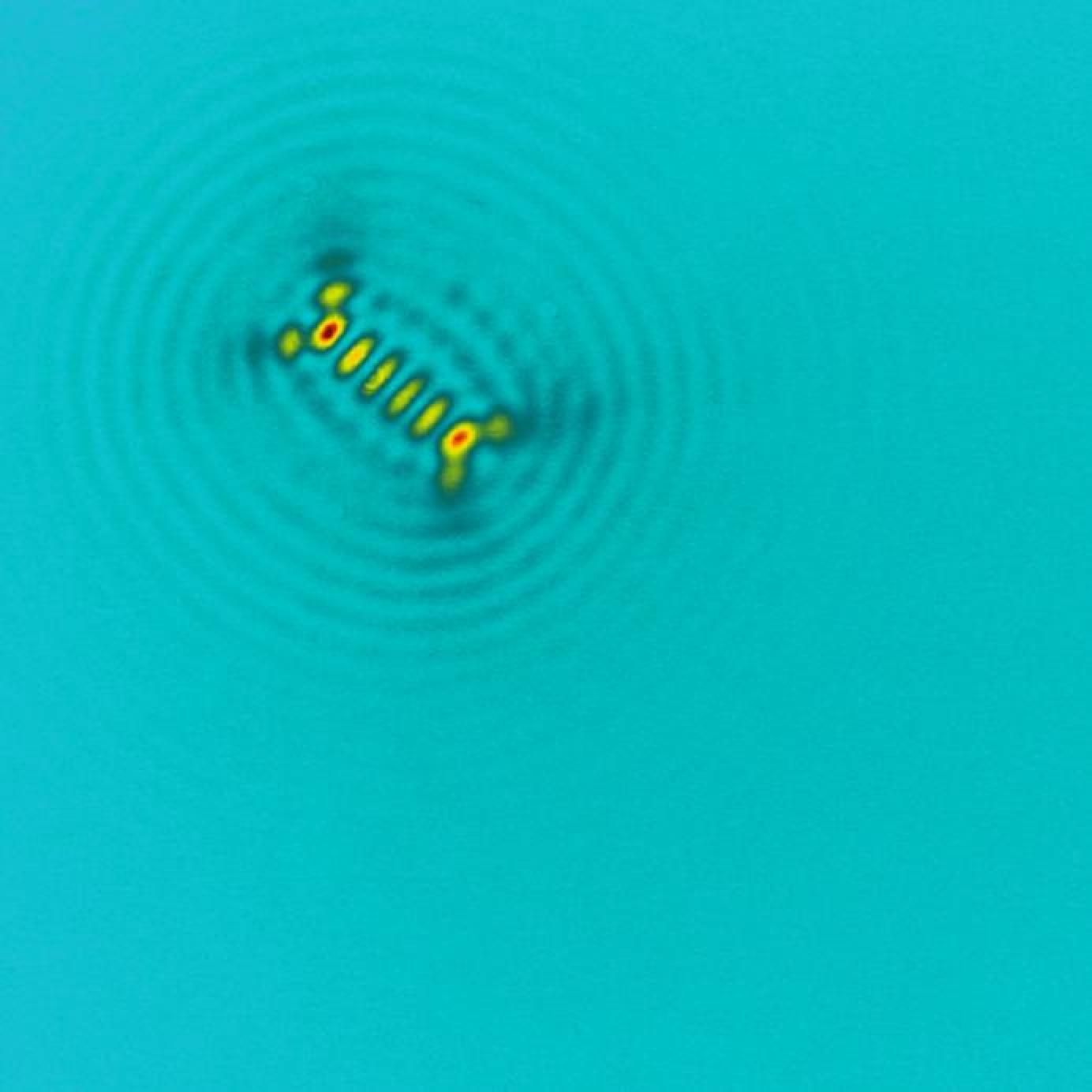
Mid-infrared laser beam profile. The central, most intense part was cut out with a "doughnut maker".
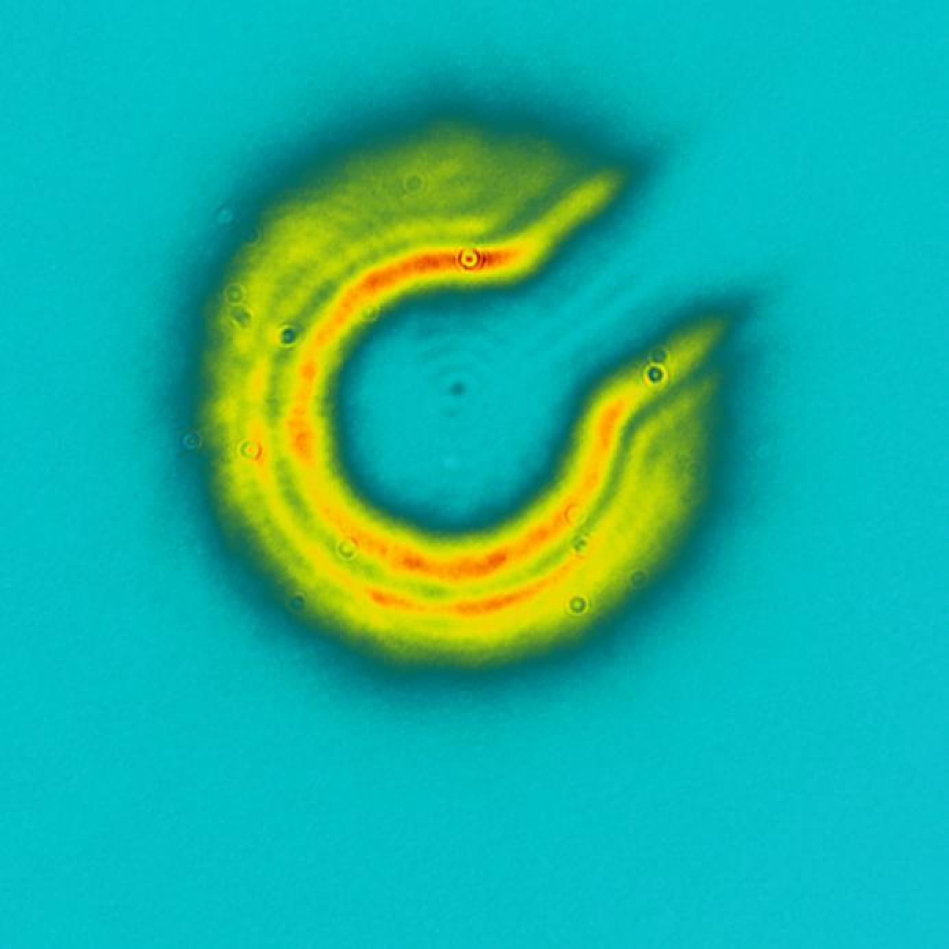
Effect of a spiral phase plate on the beam profile of a mid-infrared laser, with incorrect settings.
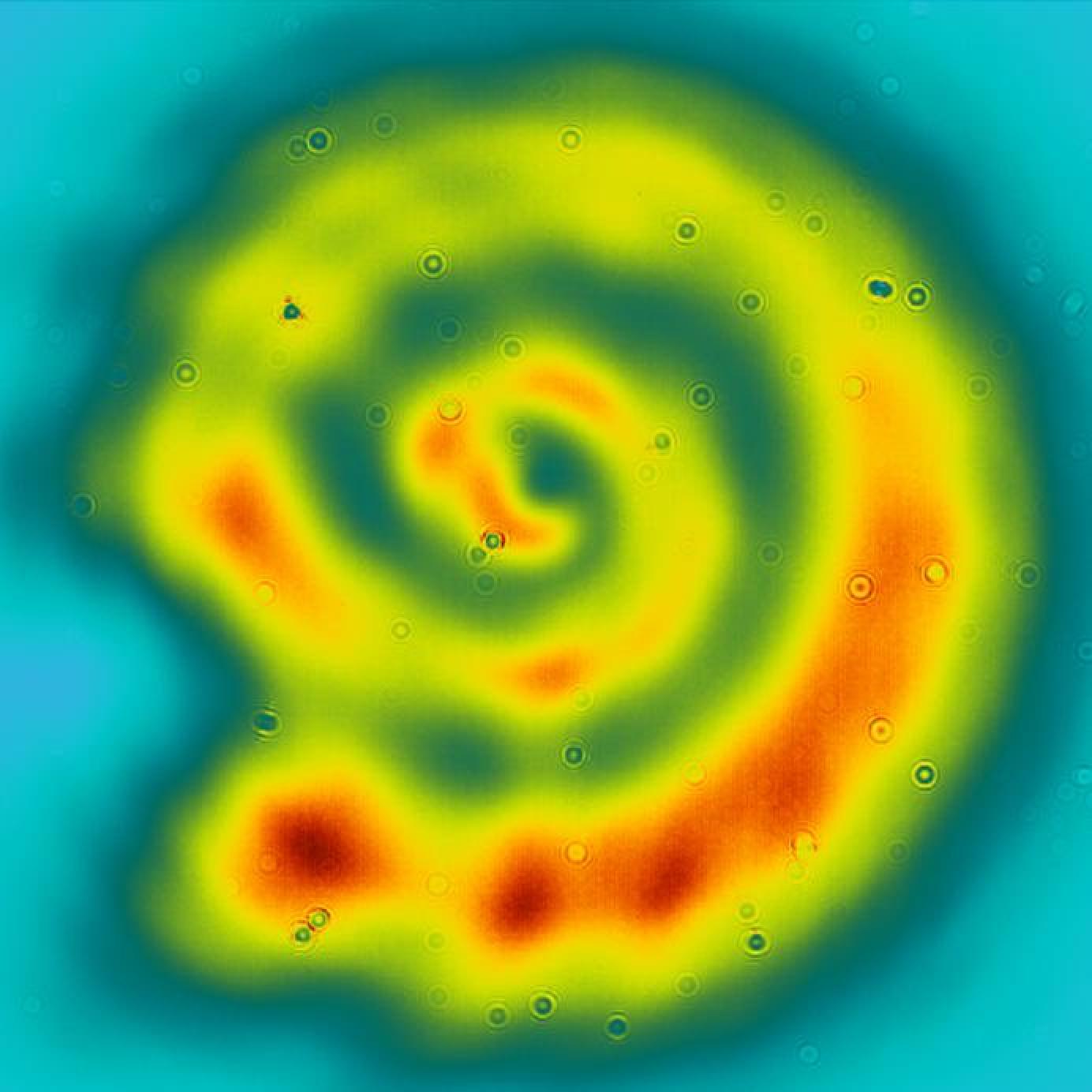
Measurement of topological charge in a structured electromagnetic field.
Mid-infrared laser beam profile with improved alignment. The central, most intense part was cut out with a "doughnut maker".
Laser induced damage on a gold-coated surface by an ultrashort femtosecond pulse in the Laser induced damage threshold end station at ELI ALPS. These damage measurements are crucial for understanding and improving optical coatings used in high-power laser facilities. Image acquired with an optical microscope at 50X magnification.
Laser induced damage on the surface of a silver mirror in LTA1.
The images to be reinterpreted can be downloaded from here.
Format requirements:
· square-shaped artworks
· minimum size 20x20 cm
Further format requirements for digital entries:
· CMYK, 300 dpi, maximum 15 MB file(s)
Information to be supplied with the entry:
· title of the artwork
· artist’s name
· artist’s age
· artist’s address (settlement)
· if relevant: name and address of the school, year, name of teacher who provided guidance for the artist
· artist’s e-mail address (in the absence of a contact e-mail we cannot notify the contestants about the results)
Deadline for the submission of entries: 7 November 2025
Digital entries must be sent to the artinscience@eli-alps.hu e-mail address, while paper based works can be left at the reception desk of the Research Institute, or sent by post to the following address: ELI ALPS Laser Research Institute, 6728 Szeged, Wolfgang Sandner u. 3. Please write “Art in Science Competition” on the envelope.
The results are planned to be announced in December 2025 at our research institute. The exact time and date will be communicated to the contestants later.
The entries will be judged by the employees of ELI ALPS. The 12 top-scoring artworks to be included in the 2026 calendar of ELI ALPS. The winners and runners-up will also receive gifts.